
Imaging membrane contact sites in live cells
Applications
A reporter that knows when to let go
Contacts between organelles form and break in seconds. splitFAST2 reports them live without holding them together, and CATCHFIRE, from the same chemistry, can act on organelles.
Why membrane contact sites are hard to image
Organelles do not work alone. They meet at membrane contact sites, places where two membranes come within roughly 10 to 30 nm of each other without fusing, and exchange calcium, lipids and signals across the gap. Nearly every organelle pair forms them, and three-way junctions exist too (Filadi et al., Nat. Cell Biol. 2026). Two properties make them difficult to watch:
- They are smaller than the resolving power of a light microscope. A 20 nm gap is far below the diffraction limit, so two organelles that look adjacent in a confocal image may be hundreds of nanometres apart. Colocalisation is not contact.
- They are transient, and the transience is the biology. Contacts form and dissolve within seconds to minutes, and cells change their behaviour by changing them. A reporter that shows where contacts are, but not when they come and go, misses the most interesting half.
The requirement is therefore unusual: a reporter that binds tightly enough to light up a 20 nm junction specifically, and loosely enough to let go the moment the junction opens. A reporter that cannot let go stops measuring the contact and starts holding it together.
Where split-GFP, FRET and engineered tethers fall short
- Split fluorescent proteins such as split-GFP reassemble into a stable barrel that does not come apart. Reviews of the field point to the “apparent irreversibility of the system” (Poggio et al., Contact 2026) and warn that such probes may stabilise contacts “possibly on the scale of days” (Filadi et al. 2026).
- FRET does not staple membranes together, but needs donor and acceptor closer than 10 nm and properly oriented, so a ratio change can reflect contact number, gap width or simply orientation (García Casas et al., Nat. Commun. 2024).
- Engineered tethers such as MAPPER deliberately hold two membranes together. That is the right tool to ask what a contact does, and the wrong one to ask when it happens.
- Proximity ligation works on fixed samples only, so the dynamics are gone before the measurement starts (Diokmetzidou and Scorrano, FEBS J. 2025).
A 2026 review concludes that low-affinity chemogenetic systems are the tools of choice to follow contact dynamics “without structural artifact” (Mukunda et al., Contact 2026).
One chemistry, two tools: observe a contact, or act on organelles
FAST is a 14 kDa protein tag with no chromophore of its own. It binds a small fluorogen and switches it on: bright when bound, dark when free, reversible at any moment. Split the tag in two, anchor one half on each membrane, and the halves reassemble only where the membranes meet, catch a fluorogen and light up the junction. When the membranes part, the signal goes. Since the first split version of 2019, the chemistry has been pushed in two opposite directions:
- splitFAST2, the lightest possible touch. The original 2019 splitFAST halves kept some affinity for each other. In 2024 the Gautier laboratory rebuilt the split tag from a FAST relative and named it splitFAST2: the two halves are about seventy times less attracted to each other (self-complementation constant of 220 µM, against 3.1 µM for the original), so they meet only because the membranes do (Rakotoarison et al., ACS Chem. Biol. 2024). Reviews list it as reversible, fast, with low self-complementation, no leaky fluorescence and second-to-minute temporal resolution (Diokmetzidou and Scorrano 2025; Poggio et al. 2026).
- CATCHFIRE, a deliberate grip. Pushed the other way, the same chemistry gives a fluorogenic molecule that does not merely reveal proximity but creates it, and lights up while it does so. Wash it out and the partners separate (Bottone et al., Nat. Methods 2023).
For reporting contacts, use splitFAST2. It is what we recommend to everyone who asks us. The papers do not always use the name: FABCON calls it splitFASTlow and PRINCESS calls it RspA-splitFAST: the same low-affinity variant.
What splitFAST2 has measured, contact by contact
ER-mitochondria contact sites and mitochondria-associated membranes (MAM)
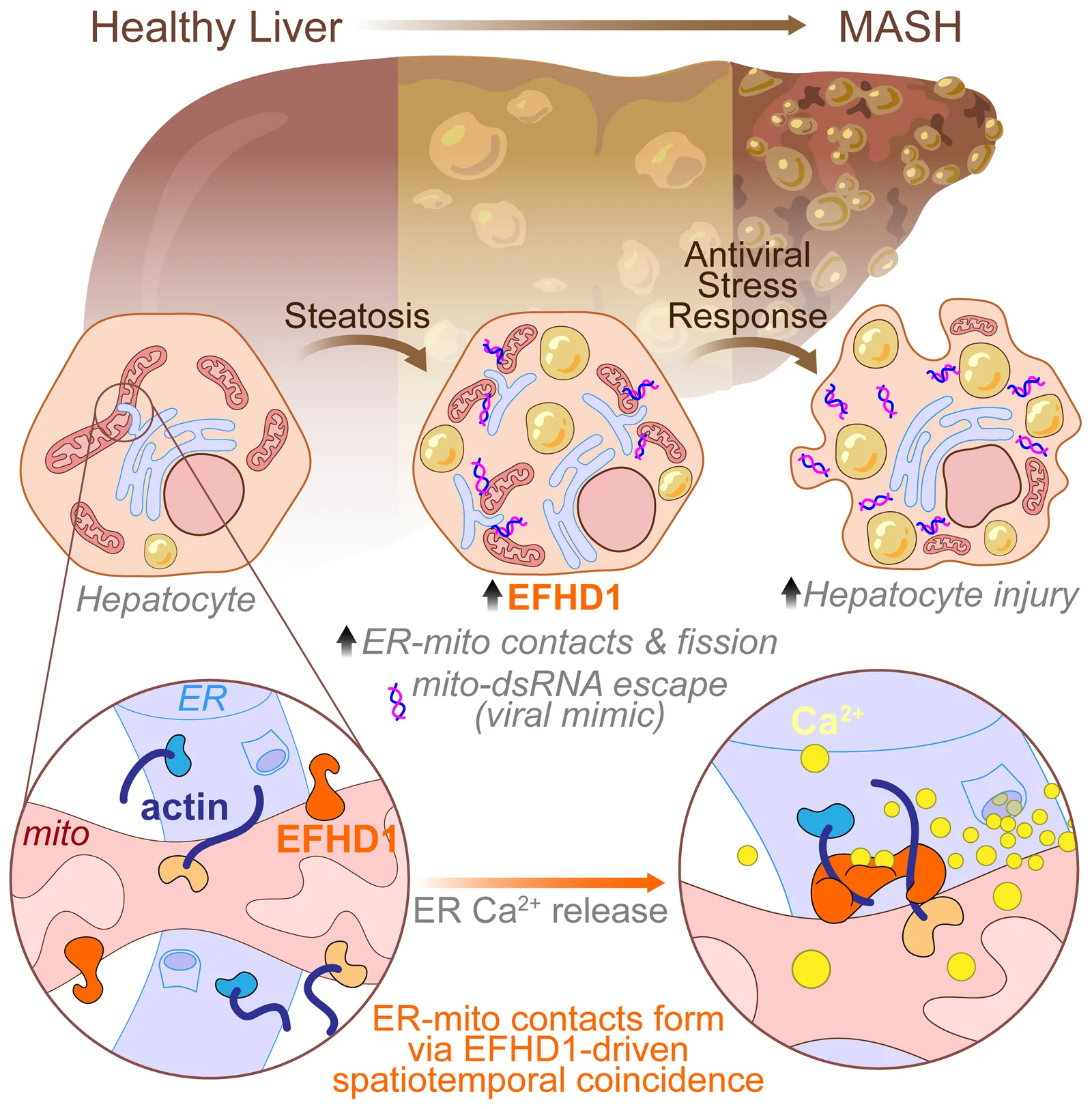
Eberhardt and colleagues asked whether the calcium-dependent actin cross-linker EFHD1 holds the ER and mitochondria together. In HepG2 liver cells, their reporter counted contacts closer than 25 nm and timed each one: in cells lacking EFHD1, far fewer fluorescent spots formed, and those that did form were shorter-lived. The study links EFHD1 to metabolic dysfunction-associated steatohepatitis (MASH) in mouse models, human liver tissue and human liver microtissues, and its authors suggest EFHD1 inhibition as a therapeutic strategy (Eberhardt et al., J. Clin. Invest. 2026; see our post on timing ER-mitochondria contacts in liver disease). That is what the tool is for: not a picture of where contacts are, but a count and a lifetime, compared between conditions.
The PRINCESS biosensors add calcium sensing to the reporter itself, so one probe reports both that a contact exists and what calcium is doing there. The same work built variants that detect contacts below 12 nm and below 25 nm, and imaged ER-mitochondria and ER-plasma membrane contacts in the same cells, told apart by colour and by fluorescence lifetime. The same probes tracked changes in ER-mitochondria contacts in four cellular models of Alzheimer’s disease: astrocytes from a transgenic mouse, astrocytes exposed to amyloid beta, neurons from an App knock-in mouse, and fibroblasts from a patient carrying a familial Presenilin 2 mutation, compared with a healthy donor (García Casas et al., Nat. Commun. 2024; see also splitFAST2 for membrane contact sites). A 2025 preprint, not yet peer reviewed, applies the published ER-mitochondria probe in mouse neuroblastoma cells carrying the familial Swedish APP mutation, a cellular model of Alzheimer’s disease (Fernandes et al., bioRxiv 2025).
Lipid droplet contacts
The FABCON toolkit built anchors for the lipid droplet surface, the ER, peroxisomes and mitochondria, then followed lipid droplet contacts as dynamic spots through droplet growth and consumption, with an automated pipeline that sorts individual contacts by size. It covers lipid droplet-ER, lipid droplet-mitochondria and lipid droplet-peroxisome contacts (Li et al., J. Cell Biol. 2024).
The lipid droplet surface is a phospholipid monolayer, not a bilayer, and the field counts droplet junctions as membrane contact sites (Filadi et al. 2026; Mukunda et al. 2026). One nuance: ER-lipid droplet junctions include membrane continuities as well as contacts, so not all of them have the two-membrane geometry of an ER-mitochondria contact.
ER-lysosome contacts, in human neurons
Vervliet and colleagues anchored one half on the ER and the other on lysosomes, with a linker sized for the 20 nm range, and showed in human iPSC-derived cortical neurons that inactive ryanodine receptors promote ER-lysosome contact formation (Vervliet et al., Nat. Commun. 2026). These neurons were not a disease model; the authors name the next step as testing whether the excessive ryanodine receptor activity seen in familial Alzheimer’s disease disturbs these contacts.
In a living animal
PRINCESS was also taken into C. elegans, with a strain reporting sarcoplasmic reticulum-mitochondria contacts in body wall muscle (García Casas et al. 2024).
When a drug changes the contacts
In two recent studies, splitFAST2 is not the subject of the work but the instrument that measures what a drug does to ER-mitochondria contacts. In rat INS-1 832/3 beta cells, ten minutes of the GLP-1 receptor agonist exendin-4 reduced the number of contacts per mitochondrial area, counted with the PRINCESS constructs, while electron microscopy showed the contacts getting shorter (Austin et al., Nat. Commun. 2025). In a preprint, not yet peer reviewed, CGP7930, a mitochondrial calcium uniporter activator found in a high-throughput screen for uniporter activity, raised the proportion of mitochondria in contact with the ER after one hour in HeLa cells, before the authors took the compound to cardiomyocytes and mouse hearts (Caldero-Escudero et al., bioRxiv 2026). One count went down, the other up: reading a fall as well as a rise takes a reporter that lets go. Both methods sections name TFLime from The Twinkle Factory as the fluorogen.
Organelle contact sites covered so far
ER-mitochondria, ER-plasma membrane, plasma membrane-mitochondria, ER-lysosome, lipid droplet-ER, lipid droplet-mitochondria, lipid droplet-peroxisome, and sarcoplasmic reticulum-mitochondria in vivo.
Which tool for which question
Ten reviews discuss splitFAST at contact sites, five of them published in 2026, and among them the field’s consensus paper on how contact sites should be defined and measured (Calì et al., Nat. Rev. Mol. Cell Biol. 2025). They converge on a division of labour rather than a single winner:
| Your question | Tool | Why |
|---|---|---|
| When does a contact form and break, in a living cell? | splitFAST2 | Reversible, live, second-to-minute resolution |
| What is calcium doing at the contact? | PRINCESS, built on splitFAST2 | One biosensor, two readouts |
| What does a contact do if I force it to exist? | An engineered tether | It makes the contact rather than reporting it |
| Does a contact exist between endogenous proteins, in fixed samples? | Proximity ligation | No overexpression, but fixed only |
| How wide is the gap? | Electron microscopy, correlative imaging | The only direct measurement of gap width |
Compiled from Diokmetzidou and Scorrano 2025, Poggio et al. 2026 and Filadi et al. 2026.
Setting up a contact-site experiment
- Distance: you detect the gap you build. The detection range is set by the linkers between each half and its membrane anchor (Diokmetzidou and Scorrano 2025). Published ER-mitochondria pairs detect contacts below 12 nm and below 25 nm, and the ER-lysosome pair was built for the 20 nm range. Choose the regime you want to interrogate.
- Readouts: number of spots and the lifetime of each (Eberhardt et al. 2026); automated classification of contacts by size (Li et al. 2024); contact and local calcium from a single biosensor (García Casas et al. 2024).
- Controls, all taken from the published work: compare a low-affinity with a high-affinity pair, the field’s own test for perturbation (Li et al. 2024); run an orthogonal assay such as proximity ligation beside the probe (Fernandes et al. 2025); include a non-expressing line or strain (García Casas et al. 2024); and where geometry matters, confirm by electron microscopy, which showed the low-affinity variant does not change contact length (Li et al. 2024).
- Limits worth knowing: fluorogen binding can lag a very fast contact event, and reassembly efficiency depends on expression level (Ke et al., Contact 2026). Keep expression modest and calibrate.
CATCHFIRE: acting on organelles
CATCHFIRE holds two tagged partners together for as long as its fluorogenic “match” molecule is present, and fluoresces while it does. It has recruited proteins to mitochondria, Golgi, ER and plasma membrane, moved lysosomes to the cell periphery and back by washout (Bottone et al. 2023), and changed cell fate in vivo by forcing unequal segregation of mitochondria at division (Bunel et al., Nat. Commun. 2025).
No CATCHFIRE contact between two organelles has been published yet. Induced tethers exist, driven by rapamycin, AP20187 or light (Ke et al. 2026). A CATCHFIRE tether would be released by simple washout, and its inducer would also be its reporter. If you are planning this experiment, we would be glad to help.
Contacts still waiting for a live reporter
Mitochondria-lysosome contacts are among the most discussed in the reviews, and no FAST-family study has reported them yet, although both anchors already exist in published FAST work (LAMP1 for lysosomes, SYNJ2BP or OMP25 for mitochondria). ER-Golgi, mitochondria-peroxisome, ER-peroxisome and endosomal contacts are also open. Three-way contacts are the other frontier: a whole 2026 review is devoted to them (Mukunda et al. 2026), and PRINCESS has already imaged two contact types at once, told apart by colour and by fluorescence lifetime.
Frequently asked
Does the reporter perturb the contact?
It depends on the generation. The original 2019 splitFAST, whose halves keep some mutual affinity (3.1 µM), stabilised ER-lipid droplet contacts in a side-by-side test; splitFAST2 (220 µM) did not (Li et al. 2024; Rakotoarison et al. 2024), and electron microscopy of mitochondria-lipid droplet contacts found that the low-affinity pair did not change their length (Li et al. 2024). This is why we recommend splitFAST2.
Are lipid droplet contacts membrane contact sites?
Yes. The droplet is bounded by a phospholipid monolayer rather than a bilayer, and the field classifies its junctions with the ER, mitochondria and peroxisomes as membrane contact sites. Some ER-droplet junctions are membrane continuities rather than appositions.
Live or fixed cells?
Live. That is the point of a reversible reporter. It has also been used in a living animal, C. elegans.
What contact distance will I detect?
The one your linkers allow. Published ER-mitochondria constructs detect contacts below 12 nm or below 25 nm, and the ER-lysosome pair was built for the 20 nm range.
Which fluorogens do I need?
splitFAST2 was characterised with fluorogens emitting between 540 and 600 nm, and the splitFAST family as a whole spans green to far-red. See the products page, and ask us which one suits your microscope and your other labels.
Disease models are described as reported by the cited studies. FAST, splitFAST and CATCHFIRE reagents are for research use only, not for diagnostic or therapeutic use.
Recent papers: splitFAST at membrane contact sites
Imaging Membrane Contact Sites Dynamics: Matching Reporter Properties to Biological Questions
A reporter-by-reporter comparison of contact site imaging, written by the SPLICS group, so by the authors of the main competing technology. split-FAST is one of four named classes and comes out of their comparative table with the shortest limitation list, a single entry, the need to add a fluorogen, against slow maturation and poor reversibility for split-GFP. Their conclusion is a division of labour rather than a ranking: fast reversible reporters such as split-FAST for resolving formation, dissociation and turnover in live cells, split-GFP for long-lived abundance and spatial organisation. They rate split-FAST in vivo applicability only moderate, which is a fair criticism. 139 references, with association and dissociation kinetics and dissociation constants given per class.
Tri-Organelle Contact Sites Integrate Metabolic Function in Health and Disease
A review of Mito-ER-lipid droplet tri-organelle contact sites. Its section 7.3 frames contact site biosensors in three generations and states that irreversible split-GFP permanently tethers the two organelles, while the low-affinity splitFAST variant used in FABCON detects contacts with minimal perturbation, confirmed by electron microscopy. Names splitFAST-based FABCON and PRINCESS as the premier tools for monitoring physiological transitions without structural artifact. No Twinkle author, no Gautier citation: independent third-party support for the non-perturbing split reporter argument.
High-throughput screen identifies a potent MCU activator boosting cardiac contractile bioenergetics
Drug discovery reaches the contact site. A high-throughput screen identifies CGP7930 as an activator of the mitochondrial calcium uniporter, binding MICU1 at a pocket between its N and C lobes, and the compound then boosts calcium-dependent mitochondrial energy metabolism in cardiomyocytes and contractile performance in mouse hearts. The contact-site measurement is a single clean experiment: HeLa cells expressing ER-Mit RspA-splitFAST with TFLime at 4 micromolar, where one hour of CGP7930 significantly raised the proportion of mitochondria in contact with the ER, which the authors read as a second mechanism for the drug beyond the uniporter itself. Preprint, not peer reviewed.
Excessive EFHD1-dependent ER-mitochondrial contacts drive a maladaptive antiviral response in metabolic liver disease
Used splitFAST revealed by TF-Lime to count ER-mitochondria contacts and time their lifetime live in HepG2 cells, showing that the Ca2+-dependent actin cross-linker EFHD1 stabilizes these contacts. In metabolic liver disease (MASH), EFHD1 excess makes contacts persist, fragments mitochondria and triggers a maladaptive antiviral stress response; inhibiting EFHD1 protects hepatocytes.
Advanced imaging of membrane contact sites
A 151-reference methods review of membrane contact site imaging, from electron microscopy and super-resolution to FRET, split-GFP/SPLICS and splitFAST. It treats reversible splitFAST reporters and PRINCESS as the way to read contact dynamics together with the Ca2+ signals they carry, and names the missing tool explicitly: no reporter yet tracks lipid transfer at contacts in real time, which the authors call a priority.
Advancing the monitoring of organelle contact sites in vitro and in vivo
A second review from the SPLICS group, Barazzuol, Peggion, Brini, Nasu and Calì, on monitoring organelle contact sites in vitro and in vivo. Same standing as Poggio: the authors of the competing split-GFP contact reporters assessing the field, with split-FAST and the full tag name both named.
Genetically Encoded Tools to Monitor and Interrogate Membrane Contact Sites
A review of the genetically encoded reporters available for membrane contact sites, split-FAST among them with thirteen mentions, set beside split-GFP and the dimerization-dependent fluorescent proteins. Published in Contact, like Mukunda, and completes the 2026 set of contact-site reporter reviews alongside Filadi and Poggio.
Inactive ryanodine receptors sustain lysosomal availability for autophagy by promoting ER-lysosomal contact site formation
Used splitFAST to study how inactive ryanodine receptors sustain lysosomal autophagy in Alzheimer's models.
Imaging and quantifying organelle contact sites using a reversible splitFAST complementation assay
A methods chapter on imaging and quantifying organelle contact sites with a reversible splitFAST complementation assay.
GLP-1R associates with VAPB and SPHKAP at ERMCSs to regulate β-cell mitochondrial remodelling and function
The GLP-1 receptor axis enters the corpus. Working in rat INS-1 832/3 beta cells, the authors show that the GLP-1 receptor associates with VAPB and SPHKAP at ER-mitochondria contact sites, and that stimulation with exendin-4 remodels mitochondria and reduces the number of contacts per mitochondrial area. The contact count is read with splitFAST2, using the PRINCESS constructs ER-RspA-NFAST and OMM-short-RspA-CFAST, in wild-type INS-1 832/3 cells only, vehicle against exendin-4 at 1 micromolar for 10 minutes, 50 to 57 cells over three independent experiments
A guide to characterizing the dynamic mitochondria-endoplasmic reticulum contact sites
A review guide to characterizing mitochondria-ER contact sites, referencing reversible splitFAST-type reporters.
Key challenges and recommendations for defining organelle membrane contact sites
The consensus paper of the contact-site field, written by twelve of its leaders including Calì and Brini at Padova, Kornmann, Hajnóczky, Rizzuto and Scorrano, and the current successor to the 2019 consensus. It defines what counts as a contact site and how one should be measured, and it treats splitFAST at length, giving the fragment boundaries, the reversibility, PRINCESS
Lipid droplet dysmetabolism affects cell homeostasis in an in vitro model of Alzheimer’s disease
Applied splitFAST to link lipid-droplet dysmetabolism to cell-homeostasis defects in an in-vitro Alzheimer's model.
You better keep an eye on your contacts
A Cell Calcium commentary written about two recent splitFAST papers
Simultaneous detection of membrane contact dynamics and associated Ca2+ signals by reversible chemogenetic reporters
Built reversible splitFAST reporters that simultaneously visualize membrane contact-site dynamics and the Ca2+ signals they carry.
Imaging and proteomics toolkits for studying organelle contact sites
A review of imaging and proteomics toolkits for organelle contact sites, including splitFAST-based approaches.
A fluorogenic complementation tool kit for interrogating lipid droplet–organelle interaction
A fluorogenic splitFAST toolkit for detecting lipid-droplet-organelle contact sites at nanometer scale in living cells.
Inter-organelle contact sites: molecular modulation and generation of new probes to study their dynamics and associated signaling
A study of inter-organelle contact sites introducing new splitFAST-related probes to visualize them.
ER-mitochondria contact sites in neurodegeneration: genetic screening approaches to investigate novel disease mechanisms
A third party recommending splitFAST rather than describing it. Reviewing the ways to see ER-mitochondria contacts in neurodegeneration, the authors set out why irreversible split reporters fall short and write that one solution would be an adapted system such as splitFAST, whose fragments dissociate when the two membranes separate.
No publications match your filters.
Other Applications

Anaerobic imaging

Detecting protein-protein interactions
Protein & cell labeling
One tag, many colors

Reversible labeling, Silent Tag
Membrane contact sites