
A reversible split reporter can tell how long an organelle contact lasts, not just whether it formed. A new study in The Journal of Clinical Investigation used splitFAST with TFLime to count contacts between the endoplasmic reticulum (ER) and mitochondria in live liver cells and to time their lifetime, second by second.
The work comes from the group of Dipayan Chaudhuri at the University of Utah, by David R. Eberhardt and colleagues, published in September 2026. The result rests on a tool that travelled through three labs. splitFAST2 was engineered in the lab of Arnaud Gautier, Twinkle’s scientific co-founder, to make its two halves less sticky. In Padova, the groups of Riccardo Filadi and Paola Pizzo built ER-mitochondria contact reporters from it, validated them and shared them through Addgene (we covered their work when it first appeared). In Utah, those plasmids answered a question about liver disease.
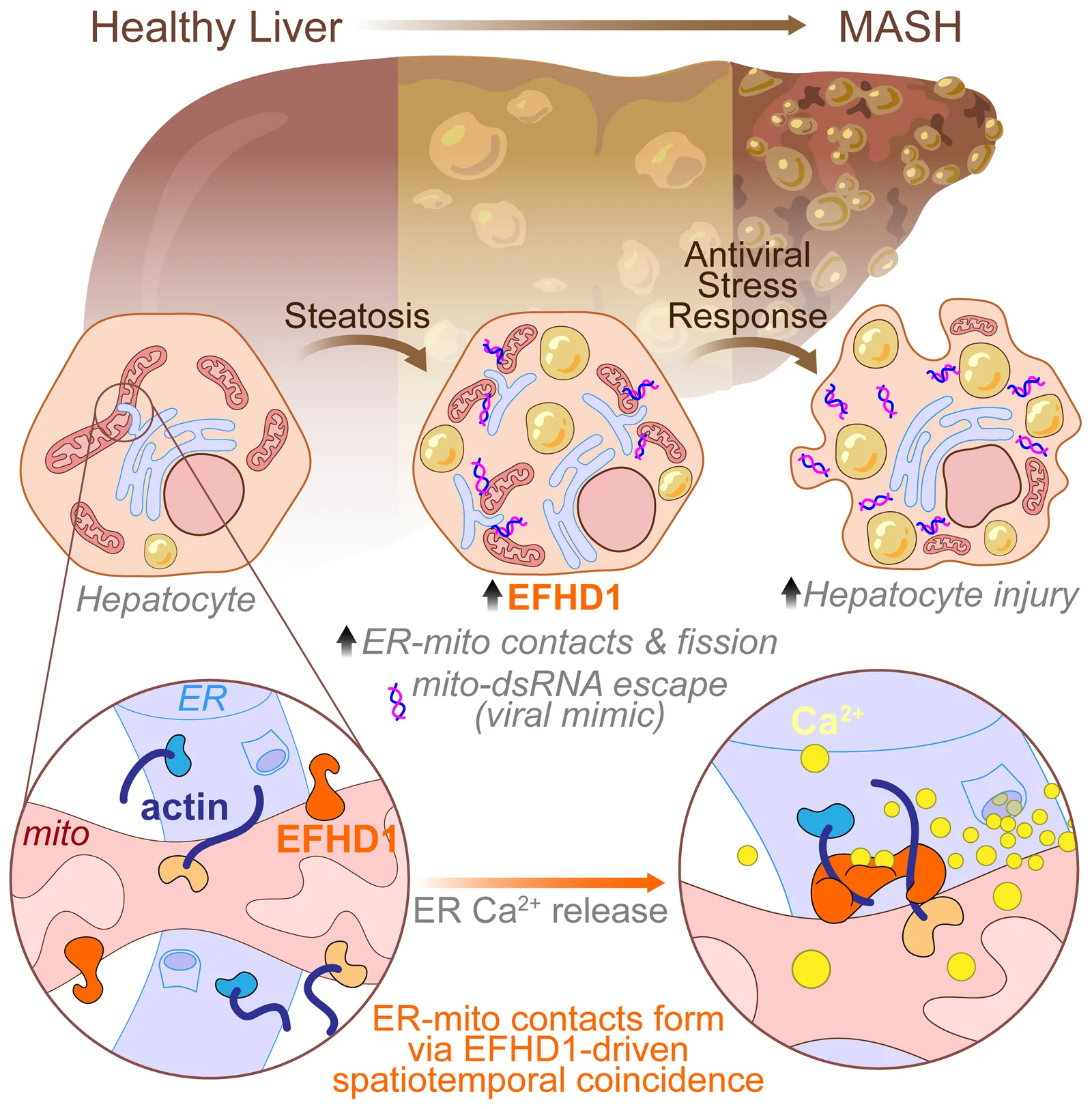
EFHD1 holds ER-mitochondria contacts together when the ER releases calcium, and too much of it drives liver injury. Graphical abstract from Eberhardt, D. R. et al., J. Clin. Invest. 2026, reproduced under a CC BY 4.0 licence. © 2026 Eberhardt et al.
A liver disease gene that sits on mitochondria
Genetic studies of liver injury, but not of liver fat, point to EFHD1, a gene of unclear function. The authors show that EFHD1 is a calcium-dependent actin cross-linker on the outer mitochondrial membrane: when the ER releases calcium nearby, it bundles actin across the gap and holds the two organelles together.
Counting contacts and timing them, live
Without EFHD1, contacts looked narrower and less regular on electron micrographs. To test their stability, the team used splitFAST: “Because splitFAST complementation is rapid and reversible, it is ideal for studying ERMCS stability.”
- The set-up. One splitFAST half sits on the ER, the other on the outer mitochondrial membrane. Where the membranes come within 25 nm, the halves assemble and light up with TFLime (5 µM).
- A reporter that watches without holding. The probes use splitFAST2 (RspA), whose halves barely attract each other. The Padova team showed by correlative light and electron microscopy that they report contacts without creating them.
- The readout. HepG2 cells were imaged every second for five minutes after an ATP pulse released ER calcium. Each spot is a contact, and it switches off when the contact ends.
- The result. Without EFHD1, contacts were far fewer and shorter-lived: 106 s on average, against 121 s under the same conditions.
An irreversible split fluorescent protein would count contacts but not time them, and would hold together the very contacts it observes.
An independent review of contact-site tools, by researchers with no connection to us, reaches the same conclusion: split GFP “permanently tethers the two organelles together”, whereas the splitFAST-based reporters “represent the premier tools to monitor true physiological transitions without structural artifact” (Mukunda et al., Contact 2026).
From contacts that last too long to liver injury
In metabolic dysfunction-associated steatohepatitis (MASH), EFHD1 rises and, the authors propose, contacts persist too long. This fragments mitochondria, releases mitochondrial double-stranded RNA and activates PKR, the liver’s antiviral kinase. Removing or knocking down EFHD1 in mice reduced liver damage, inflammation and fibrosis, and human genetics supports the same chain. The authors propose EFHD1 as a target to uncouple liver injury from fat accumulation.
For contact-site biology, a contact’s lifetime is a variable in its own right, and a reversible reporter can measure it in living cells.
Set it up in your cells
- Reporters. ER-long RspA-NFAST and OMM-short RspA-CFAST, from the Filadi lab, available through Addgene (233600 and 233594).
- Fluorogen. TFLime at 5 µM, green channel (488 nm excitation, 520 nm emission), compatible with a far-red mitochondrial stain.
- Analysis. Track spot intensity over time and count a contact while its normalised intensity stays above half its maximum.
More reading on splitFAST at organelle contacts
- Excessive EFHD1-dependent ER-mitochondrial contacts drive a maladaptive antiviral response in metabolic liver disease. Eberhardt, D. R. … Chaudhuri, D. J. Clin. Invest. 2026, 136(19), e204023. doi.org/10.1172/jci204023
- Simultaneous detection of membrane contact dynamics and associated Ca2+ signals by reversible chemogenetic reporters. García Casas, P. … Filadi, R. Nat. Commun. 2024, 15, 9775. doi.org/10.1038/s41467-024-52985-0
- Improving Split Reporters of Protein-Protein Interactions through Orthology-Based Protein Engineering. Rakotoarison, L. M. … Gautier, A. ACS Chem. Biol. 2024, 19(2), 428-441. doi.org/10.1021/acschembio.3c00631
- Tri-Organelle Contact Sites Integrate Metabolic Function in Health and Disease. Mukunda, S. G. … Katti, P. Contact 2026, 9. doi.org/10.1177/25152564261496636
- Imaging and quantifying organelle contact sites using a reversible splitFAST complementation system. Weesner, J. A., Chang, C. L. Methods Enzymol. 2026, 75-91. doi.org/10.1016/bs.mie.2025.11.023
Recent Comments