
A chemical inducer can decide what a protein pair is made of, and that turns a question about stoichiometry into a picture. A group in Aachen used CATCHFIRE to assemble STAT5 pairs containing one working and one mutated protomer, and found that a single working copy of the import motif they identify is enough to carry the pair into the nucleus.
The work comes from Sabrina Ernst and Gerhard Müller-Newen at the Institute of Biochemistry and Molecular Biology, Uniklinik RWTH Aachen, published in Communications Biology on 26 September 2026. It is peer reviewed and open access. CATCHFIRE was developed in the lab of Arnaud Gautier, Twinkle’s scientific co-founder. What the technology contributes here is not a picture of where a protein sits, but control over which two proteins are joined.
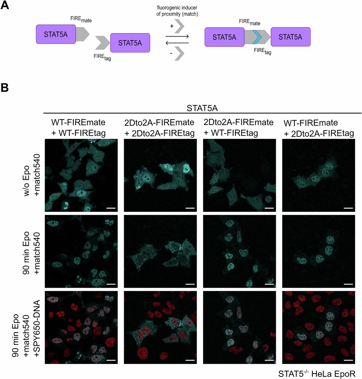
Assembling a STAT5 pair of chosen composition. FIREmate on one protomer and FIREtag on the other, joined on addition of match540 (top). Four wild-type and mutant combinations in STAT5-deficient HeLa cells with match540 alone, where the signal is mostly cytoplasmic, then after 90 minutes of erythropoietin, then with a nuclear stain added (bottom). These are representative images rather than a quantification. Reproduced from Figure 8 of Ernst, S. et al., Commun. Biol. 9, 1257 (2026), under a CC BY 4.0 licence. © The Author(s) 2026.
A transcription factor that cannot get in
The transactivation domain switches STAT5’s target genes on, and was not thought to take part in the travelling. Yet a STAT5A mutant lacking it is phosphorylated with the same kinetics as the wild type and dimerizes normally after erythropoietin, then accumulates in the cytoplasm instead of the nucleus. The equivalent STAT3 mutant does the opposite, so this is not a family-wide rule. Twelve residues carry the effect, and within them two acidic residues, D754 and D758, are essential. The authors read them as part of an address that exists only once two protomers are together.
Building the dimer you want to test
- Why co-expression cannot answer it. If the address belongs to the pair, does the pair need it twice? Putting a working and a broken version in the same cell gives three kinds of pair, and the microscope cannot say which one it is watching.
- The design. FIREmate was fused to one STAT5A protomer and FIREtag to the other, in four constructs. Adding match540 joins one of each, so the composition of the assembled pair follows from which two constructs are in the cell.
- The result. Pairs made of one wild-type and one mutated protomer accumulated in the nucleus, in both tag orientations. One intact domain per pair is enough.
- What the inducer did not do. On its own match540 was no substitute for the cytokine. Forced wild-type pairs became nuclear in a subset of cells only, and complete import across all cells needed erythropoietin.
- A detail worth copying. The inducer reported its own assembly, so the cells carrying both constructs were identified by the fluorescence that appeared when it was added.
The FIREmate and FIREtag plasmids came from us, as the methods state. Our reagents are sold for research use only.
Why it matters for myeloproliferative neoplasms and leukemia
Persistent or dysregulated STAT5 activation drives myeloproliferative neoplasms and leukemia, the authors note. Their link to that biology is measured rather than asserted: in cells driven by BCR-ABL, the oncogenic kinase of chronic myeloid leukemia, a STAT5A lacking the whole domain that carries the motif also stayed in the cytoplasm, and the authors read that as the motif being required there too. On that basis they propose the motif as a potential target for therapeutic intervention. The work is in cell lines. There is no animal model, no patient sample and no molecule that blocks the motif, and the authors put the next step in the future tense themselves.
More reading
- Unexpected functional role of the transactivation domain for nuclear import of STAT5. Ernst, S., Borgen-Möller, M., Küster, A., Schurse, H., Müller-Newen, G. Commun. Biol. 2026, 9, 1257. doi.org/10.1038/s42003-026-10898-2
- A fluorogenic chemically induced dimerization technology for controlling, imaging and sensing protein proximity. Bottone, S. et al. Nat. Methods 2023, 20 (10), 1553-1562. doi.org/10.1038/s41592-023-01988-8
- Chemically Responsive Protein Switches for the Precise Control of Biological Activities. Franco Pinto, J. et al. ACS Chem. Biol. 2026, 21 (8), 1958-1971. doi.org/10.1021/acschembio.6c00360
- What the same chemistry does when the point is to act rather than to observe: reversible protein switches with CATCHFIRE, and the mechanism itself on the science behind CATCHFIRE.
Recent Comments