A split reporter can tell whether a protein complex has been built, cell by cell. A preprint from Harvard uses splitFAST with TFLime to read out the assembly of cytochrome c oxidase (complex IV) in the mitochondria of living yeast, and the readout helps explain why genetically identical cells settle into two different ways of making energy.
The work comes from Piyush Nanda and Andrew W. Murray at Harvard University, posted on bioRxiv in March 2026 and revised in September. It is a preprint and has not yet been peer reviewed. splitFAST was engineered in the lab of Arnaud Gautier, Twinkle’s scientific co-founder. FAST has been used in budding yeast for several years. What is new is the question it answers: not where a protein is, but whether a complex has assembled.
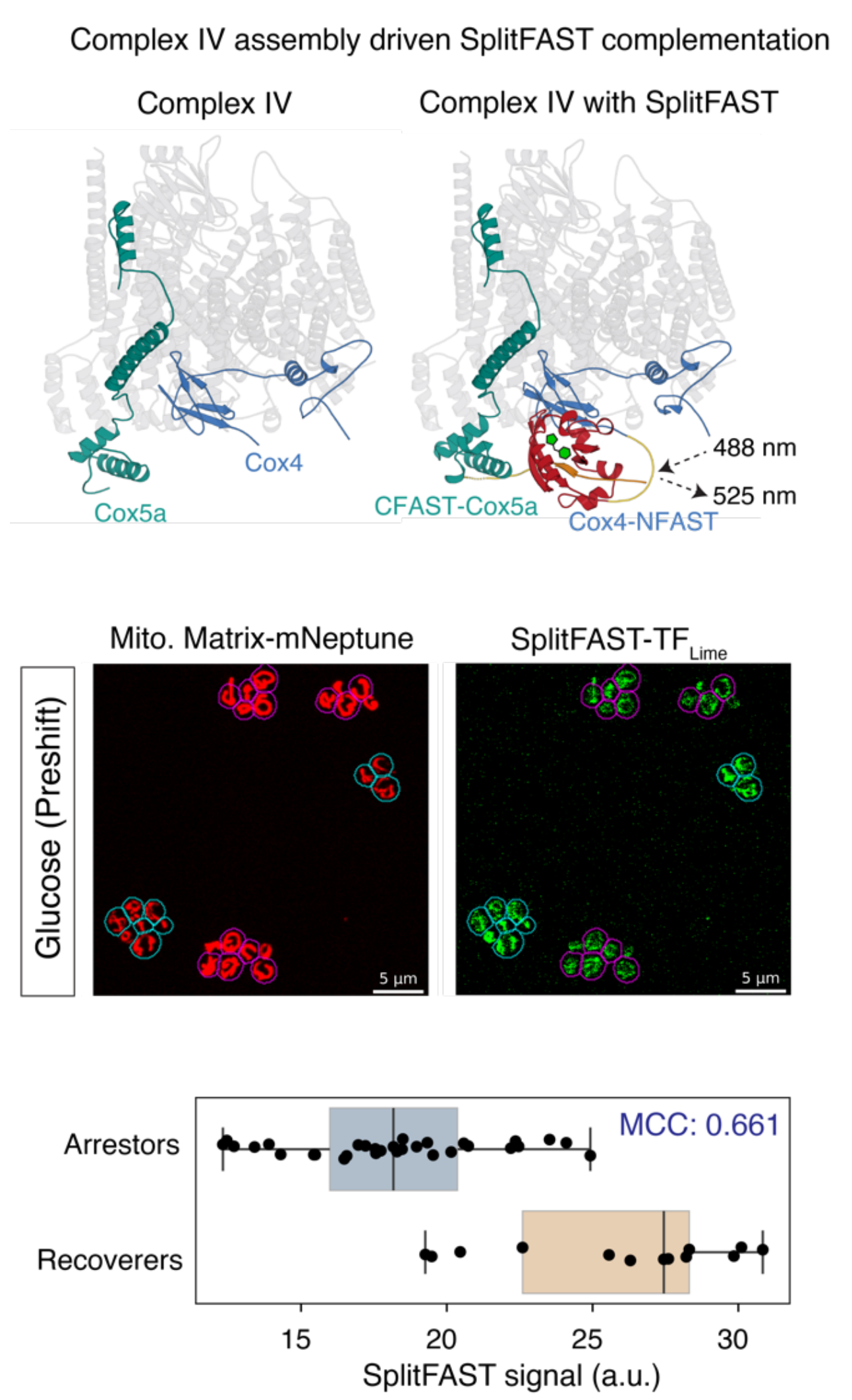
splitFAST as a complex IV assembly reporter. The two halves meet only in the assembled complex (top). Matrix marker and splitFAST-TFLime signal in yeast on glucose, cyan outlines for recoverers, magenta for arrestors (middle). Signal per cell in the two states (bottom). Adapted from Figure 3 E-G of Nanda, P. & Murray, A. W., bioRxiv 2026 (preprint), under a CC BY 4.0 licence. © 2026 the authors.
Two kinds of cells in the same culture
Yeast growing on glucose holds two kinds of cells. Arrestors ferment and stall when glucose runs out. Recoverers respire and resume growth on other carbon sources. The Murray lab described this bet-hedging in 2020. The preprint proposes what holds each cell in its state: a feedback loop in which the mitochondrial membrane potential drives the import of mitochondrial ribosome proteins, these ribosomes make three core subunits of complex IV, and complex IV sustains the potential. To test that loop, the authors needed to measure how much complex IV each cell had assembled.
Reading out complex IV assembly with splitFAST
- The design. Guided by the structure of yeast complex IV, the authors fused NFAST to Cox4 and CFAST to Cox5a at their genomic loci. The two subunits sit side by side only in the assembled complex, so that is where the halves complement and light up with TFLime.
- The controls. The signal was highest with both halves present, and fell when the structural subunit Cox6 or the assembly chaperone Pet191 was deleted.
- The control that matters most here. Arrestors and recoverers differ in membrane potential, so a fluorogen whose entry followed the potential would create a difference on its own. The authors dissipated the potential before adding the fluorogen, and the signal did not drop.
- The result. Recoverers carry more assembled complex IV than arrestors, consistent with their higher oxygen consumption.
- A quantitative check. When mitochondrial translation was dialled down with chloramphenicol, the splitFAST signal fell with a Hill coefficient of 3.45 ± 0.28, close to the 3 the authors predicted and to the 3.05 they measured from oxygen consumption.
A fluorescence readout taken in single living cells thus gave the same cooperativity as a bulk measurement of the complex’s activity.
Why it may matter beyond yeast
The authors propose that mitochondrial and cytoplasmic translation compete. Reducing mitochondrial translation recreated the two states in fission yeast, which normally shows only one. In the preprint’s discussion, they speculate that the same competition could help explain the Warburg effect, the fermentation of cancer cells in the presence of oxygen. These are the authors’ proposals in a preprint and may change with peer review.
Set it up in your cells
- Constructs. Cox4-(GGGGGS)2-NFAST and Cox5a with CFAST10 after its mitochondrial targeting sequence, tagged at the native loci by CRISPR-Cas9. The authors invite requests for strains.
- Fluorogen. TFLime, green channel (488 nm excitation, 525 nm emission). The preprint does not give the concentration.
- Imaging. Spinning-disk confocal, 60x/1.4 NA, z-stacks through the whole cell, signal normalised to a mitochondrial matrix marker.
- A control worth copying. If your conditions change the membrane potential, dissipate it before adding the fluorogen to show that the signal reports assembly, not uptake.
More reading
- Translational antagonism enables bet-hedging in yeast via mitochondrial bistability. Nanda, P., Murray, A. W. bioRxiv 2026 (preprint). doi.org/10.64898/2026.03.30.715437
- A Putative Bet-Hedging Strategy Buffers Budding Yeast against Environmental Instability. Bagamery, L. E. … Murray, A. W. Curr. Biol. 2020, 30, 4563-4578. doi.org/10.1016/j.cub.2020.08.092
- A split fluorescent reporter with rapid and reversible complementation. Tebo, A. G., Gautier, A. Nat. Commun. 2019, 10, 2822. doi.org/10.1038/s41467-019-10855-0
- splitFAST in mitochondria, the other way round: timing ER-mitochondria contacts in liver disease.
Recent Comments