
Inside an actin filament, a nucleotide stays put until its subunit leaves. In MreB, the bacterial relative of actin, it does not, a new preprint reports. Using pFAST to watch single filaments on a lipid bilayer, the authors saw nucleotide being exchanged along filaments already formed.
The work comes from the group of Rut Carballido-López at the Micalis Institute (INRAE, Université Paris-Saclay), with first author Ingrid Adriaans. It was posted as a preprint on Research Square in July 2026. “To our knowledge, this is the first report of nucleotide cycling within a biological polymer,” the authors write. They acknowledge Arnaud Gautier, Twinkle’s scientific co-founder, for pFAST tag and fluorogens.
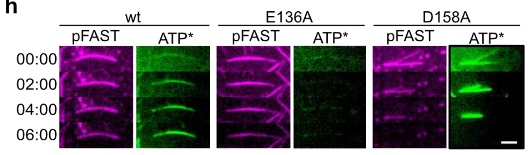
Fluorescent ATP (green) loading along preformed pFAST-MreB filaments labelled with TFCoral (magenta). Wild type takes it up, E136A does not (green signal enhanced to detect it), D158A is about twice as fast. Scale bar, 2 µm. Reproduced from panel h of Figure 3 of Adriaans, I. E. et al., Research Square (2026), preprint, under a CC BY 4.0 licence. © The Author(s) 2026.
What the filaments did
- ATP binding builds them. As in actin, hydrolysis comes later, within the filament. Two mutants with markedly reduced ATPase activity still polymerised.
- Nucleotide reaches the inside. Preformed filaments took up fluorescent ATP along their length, over minutes. This is where MreB parts company with actin.
- Free nucleotide decides their fate. In excess ATP, filaments stayed stable for up to several hours. Flushed with ADP, they vanished in under a minute. In nucleotide-free buffer, they took about 20 minutes.
What pFAST did here
Filament dynamics are known to be very sensitive to tagging, and MreB is “notoriously difficult” to purify and label. A pFAST fusion let the authors follow single filaments in real time by TIRF microscopy. TFLime carried the imaging and the elongation kinetics. TFCoral took over when fluorescent ATP needed the green channel: same tag, new colour.
And pFAST passed the authors’ checks. Tagged and untagged MreB form the same filament pairs. The growth-rate plateau agrees with label-free high-speed atomic force microscopy, a sign, they write, that it “is not an artefact of the pFAST tag or its labelling strategy”. Under ADP, untagged filaments disassemble similarly. And the fusion, they report, is fully functional in vivo.
They used TFLime and TFCoral, our fluorogens (research use only).
What’s next
Next comes imaging single filaments inside living cells, where MreB so far appears as diffraction-limited patches. A first step is already there: in Bacillus subtilis expressing GFP-MreB, ATP depletion delocalised wild-type MreB but not the E136A mutant. The authors propose that high ATP keeps filaments on the membrane in growing cells, and that the ATP/ADP ratio may control MreB. Sporulation, stationary phase and stress are natural places to look.
More reading
- Intrafilament nucleotide exchange in a prokaryotic actin homolog. Adriaans, I. E. … Carballido-López, R. Research Square 2026 (preprint). doi.org/10.21203/rs.3.rs-10303285/v1
- Engineering of a fluorescent chemogenetic reporter with tunable color for advanced live-cell imaging. Benaissa, H. … Gautier, A. Nat. Commun. 2021, 12, 6989. doi.org/10.1038/s41467-021-27334-0
Recent Comments